The X-ray Tube:
X-ray tubes are found in a wide variety of imaging devices, Computed Tomography (CT), portable x-ray machines, mammography machines, fixed position x-ray machines, fluoroscopy units, and c-arm x-ray machines.
Medical x-rays are produced in a glass enclosed vacuum tube. (See Figure 2.1 and 2.2) A high voltage differential is applied across a gap in a vacuum tube between a cathode and an anode. The anode is the target for the electrons that are accelerated across the vacuum and the anode is usually made of a metal, often tungsten. When the voltage differential results in an electron crossing over the gap and impacting the metallic anode the electron slows down and liberates energy as heat and x-rays. The majority of the energy is liberated as heat. This type of interaction that results in x-ray generation, has been called by the “braking of the electron”, the bremsstrahlung effect.
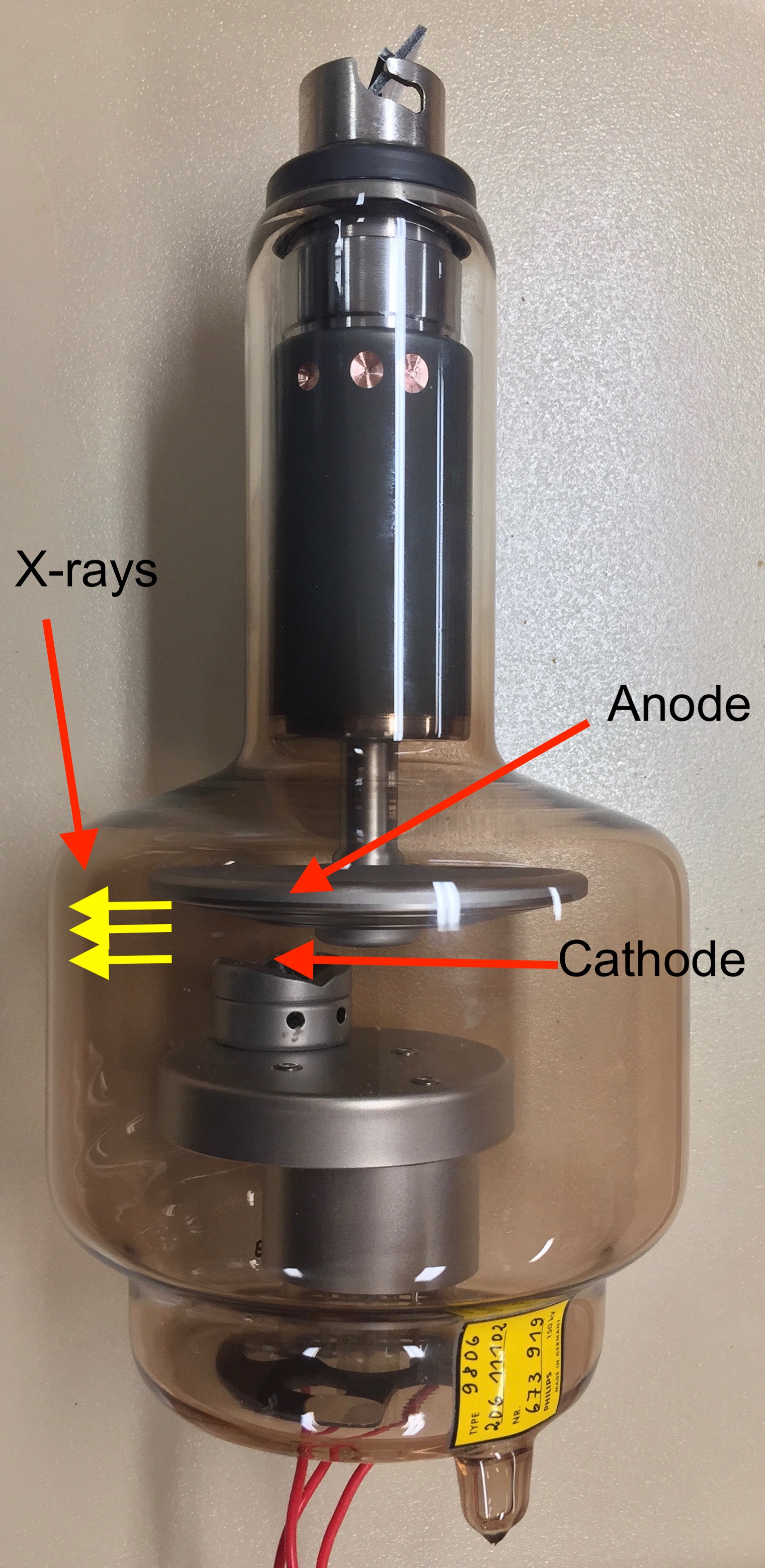
Fig 2.1) This image demonstrates a full-length view of a glass x-ray tube with the cathode and anode illustrated.
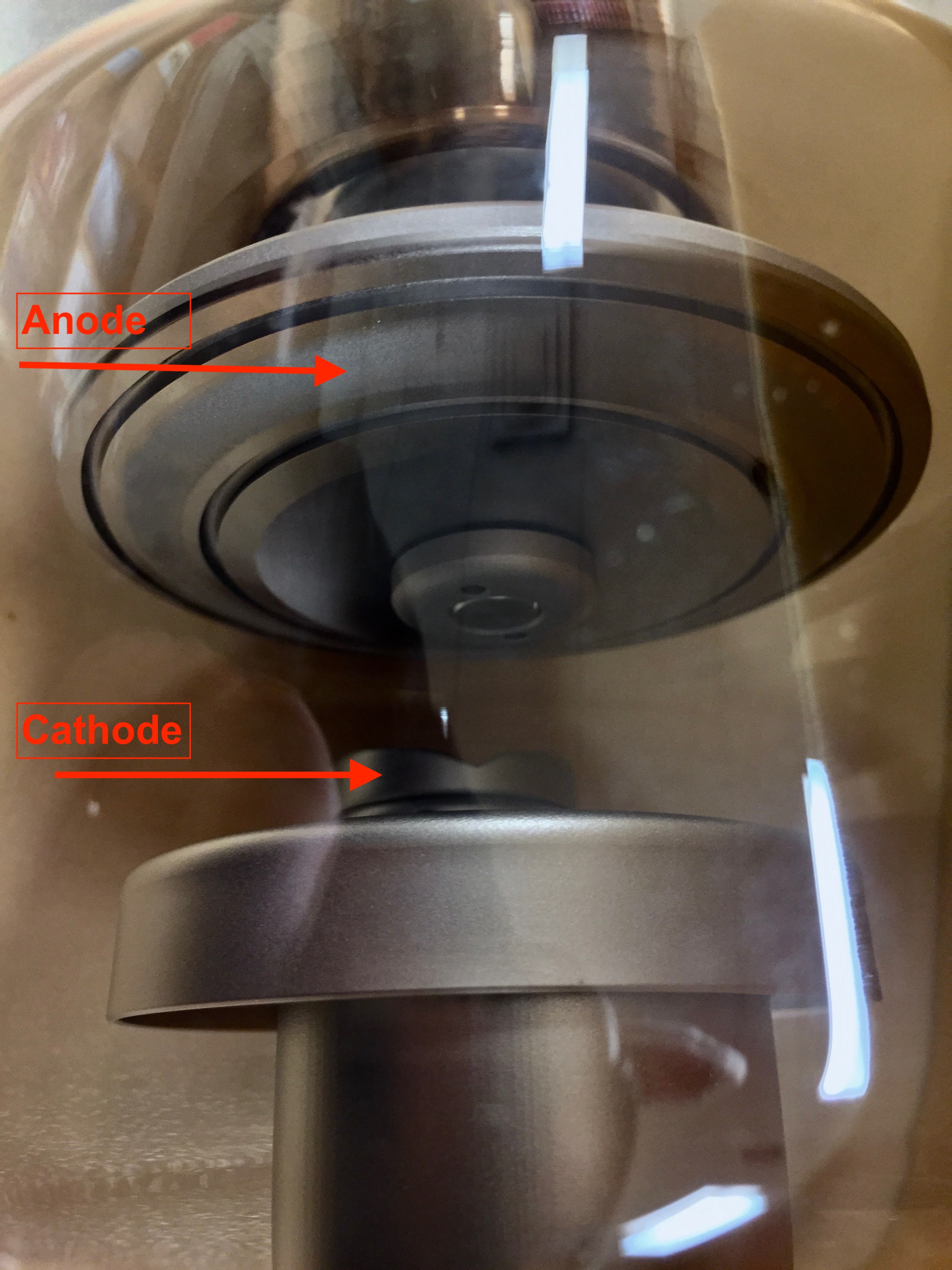
Fig 2.2) This magnified image focuses upon the relationship between the cathode and the anode.
X-ray tubes are usually associated with apparatus that allows for the physical positioning of a patient for an examination and an x-ray detection system that collects transmitted x-rays and transforms them into images. (Figure 2.3)
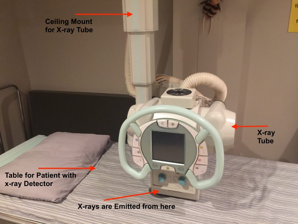
Fig. 2.3) A ceiling mounted x-ray tube is seen. The x-rays are collimated and emitted from the end of the housing that is resting on the x-ray table. The x-ray detector is below the surface of the table.
The x-ray tube has physical features that allow for the cooling of the components related to the heat produced and for focusing and directing of the x-rays towards the target (the patient). The x-rays created are collimated by the lead housing of the x-ray tube and by mobile metallic plates in the x-ray tube which results in the x-rays being emitted from a small aperture in the x-ray tube housing. (Figure 2.4)
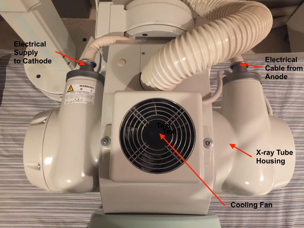
Fig. 2.4) This is an overhead view of the same x-ray tube seen in Figure 2.3. The electrical cables associated with the cathode and the anode are seen. The glass x-ray tube is inside the lead housing. The cooling fan is seen projecting from the top of the x-ray tube housing.
The x-rays emitted from the x-ray tube have three possible fates when encountering human anatomy:
a) They are transmitted through the patient to interact with the x-ray detector on the opposite side of the entry site, resulting in an image;
b) They are absorbed by the patient’s tissues and the energy is dissipated;
c) They are scattered by the patient’s tissues and leave the body in another direction from the entry site. These scattered x-rays may interact with the detector or they may enter the physical space surrounding the patient.
Different tissues absorb x-rays differently based upon their molecular structure i.e. bone vs. fat and upon the density and the thickness of the tissue. This differential absorption of the incident x-ray beam results in the differential detection of the x-ray beam after exposure of the detector and hence is directly responsible for the varying intensities seen on the gray scale of the resulting image.